neurology

Episode 41 – Vertigo

(ITUNES OR LISTEN HERE)

The Free Open Access Medical Education (FOAM)

We cover two bits of FOAM, one from Emergency Medicine Literature of note on the use of meclizine for vertigo and an EMcrit episode on the HiNTs exam.

Emergency Medicine Literature of Note – Dr. Ryan Radecki – Treating what you believe is peripheral vertigo?  Using meclizine? So are most people.  But this is not an evidence based practice.  While meclizine is Rosen approved, Tintinalli recommends transdermal scopolamine as the first line treatment [1,2]. Following a recent recall of meclizine (oddly because iron bottles contained meclizine instead of iron), Dr. Radecki probes into why we use meclizine for vertigo.

  • Meclizine is an anti-histamine and has been thought to have anti-emetic properties.
  • A 1968 paper compared 16 anti-emetics/combinations and did NOT conclude that meclizine was the best. In fact, scopolamine and amphetamine performed best. Promethazine (phenergan) is also a good choice based on this paper [3].

EMcrit – Dr. Scott Weingart – The HiNTs exam has taken off, particularly in the FOAM world, as a means of disguising between central and peripheral causes of vertigo. In 2010, an EMcrit episode popularized this in the FOAM world. See this video demonstrating the HiNTs exam.

  • Head Impulse – rapid head rotation by the examiner with the subject’s vision fixed on an object (i.e. examiner’s nose). The examiner rapidly rotates the head towards midline and the patient’s eyes should remain fixed on the target. 
    • Abnormal (loss of fixation on target and movement of eyes away from target followed by correct saccade as patient fixates on target) = peripheral (vestibulo-ocular reflex impaired)
    • Normal = central
  • Nystagmus 
    • Horizontal nystagmus with a unidirectional fast phase (away from affected side) = probably peripheral
      • Patients with horizontal nystagmus may have central pathology but may have direction-changing nystagmus (i.e. fast phase beating in one direction when looking to right and the opposite direction when looking left).
    • Vertical or nystagmus = central pathology
  • Test of Skew Patients should fixate on a target while the provider alternately covers each eye. As the cover is moved from one eye to the other, the uncovered eye must correct for the misalignment and will look up or down to focus back on the target. This slight correction is observed repeatedly as the cover is moved from one eye to the other.
    • Skew deviation/misalignment = probably central, often in posterior fossa abnormalities
    • No skew deviation= peripheral

INFARCT – Impulse Normal, Fast-phase Alternating nystagmus, and Refixation on Cover Test

Issues with HiNTs

  • Can only be performed on patients with continuous vertigo.
  • External validity is a major issue with HiNTs.
    • Providers – Of the 4 studies have examined the operating characteristics of HiNTs, none have used emergency providers and instead have examined how the exam performs in the hands of two neuro-ophthalmologists, neuro-otologists, and neurologists with 4 hours of specialized training in the exam. It’s unclear whether HiNTs would be reliable or valid when performed by emergency providers [4-6].
    • Patients – The patients examined in many of these studies have other indicators of badness on neurologic exam. In one study, patients had to have gait instability and/or truncal ataxia to enroll.  Then, 76/101 (76%) of those patients had a central cause. These patients were sick and not the undifferentiated vertiginous patients we see primarily as emergency providers [4-6].
    • In the words of leading HiNTs expert Dr. Newman-Toker, HiNTs “requires expertise not routinely available in emergency departments.” As such, his team is piloting quantitative video-oculography to aid in diagnosis using HiNTs [7]. An Annals of Emergency Medicine review also warned that HiNTs may not be ready for emergency provider use [9].

More FOAM on HiNTs: EMJclub, EMNerd

Core content 

We delve into core content on vertigo using Rosen’s Medicine (8e) Chapter 19,  and Tintinalli’s Emergency Medicine: A Comprehensive Study Guide  (7e) Chapter 164 “Vertigo and Dizziness.”

Vertigo is often characterized by the sensation of spinning and falls into the broad and frustrating category of “dizziness.”  Often, when a dizzy patient presents we perseverate on characterizes what the patient means by “dizzy.” However, some argue that this is not an appropriate approach as a study found 50% of patients changed the character of their dizziness when questioned again after 10 minutes [9].  Additionally, the clinical characteristics differentiating peripheral from central causes of vertigo are not entirely reliable. Despite these limitations, it is expected that we are familiar with “classic” presentations.

Screen Shot 2016-01-13 at 8.32.04 AM

*”Classic” presentations

Generously Donated Rosh Review Questions 

  1. A 50-year-old man presents with episodic severe vertigo lasting hours, with associated symptoms of unilateral tinnitus, fluctuating low-frequency hearing loss, and aural fullness. 
  2. A 20-year-old woman presents with an acute onset of dizziness. The patient describes the sensation that the room is spinning when she turns her head to the left and it is accompanied by nausea and vomiting. The symptoms resolve with turning her head away from that side. Examination reveals nystagmus elicited by deviating the eyes to the left and no other neurologic findings. 

Answers

1. B. Meniere’s disease is characterized by episodic severe vertigo lasting hours, with associated symptoms of unilateral tinnitus, fluctuating low-frequency hearing loss, and aural fullness. Typical onset is in the fifth decade of life. The cause is uncertain but is speculated to result from allergic, infectious, or autoimmune injury. The histopathologic finding includes endolymphatic hydrops, which is thought to be caused by either overproduction or underresorption of endolymph in the inner ear. Meniere’s disease is a clinical diagnosis mostly based on history. Testing may be obtained to support the diagnosis and rule out other disorders. Audiometry often demonstrates a low-frequency sensorineural hearing loss. An FTA-ABS test may be obtained to rule out syphilis. Electronystagmography (ENG) may demonstrate a unilateral peripheral vestibular weakness on caloric testing. When the diagnosis is uncertain, a brain MRI with contrast is obtained to evaluate for a retrocochlear lesion. The differential diagnosis of Meniere’s disease includes acute labyrinthitis, neurosyphilis, labyrinthine fistula, autoimmune inner ear disease, vestibular neuronitis, and migraine-associated vertigo.The most common cause of peripheral vestibular vertigo in adults is benign paroxysmal positional vertigo (A). BPPV occurs in all age groups but more often between ages 50 and 70 but is not associated with hearing loss and made worse with movement. In a perilymph fistula (C) rapid changes in air pressure (barotrauma), otologic surgery, violent nose blowing or sneezing, head trauma, or chronic ear disease may cause leakage of perilymph fluid from the inner ear into the middle ear and result in episodes of vertigo. Associated signs and symptoms are variable but can include a sudden pop in the ear followed by hearing loss, vertigo, and sometimes tinnitus. Acute vertigo associated with nausea and vomiting (but without neurologic or audiologic symptoms) that originates in the vestibular nerve is known as vestibular neuronitis (D). Vestibular neuronitis can occur spontaneously or can follow viral illness.

2. B. This patient presents with peripheral vertigo most consistent with benign paroxysmal peripheral vertigo (BPPV) and should be treated with an Epley maneuver. Vertigo is defined as the sensation of disorientation in space combined with a sensation of motion. Patients typically describe a “room-spinning” sensation or the feeling of sea sickness. Vertigo can be divided into two types: central and peripheral. Central vertigo are those disorders arising from the central nervous system and include ischemic stroke, vertebrobasilar insufficiency and infectious causes (meningitis, mastoiditis, syphilis). Central vertigo is characterized by longer duration of symptoms, minimal change with position, gradual onset and multidirectional nystagmus. Peripheral vertigo includes BPPV, Meniere’s disease, Labyrinthitis and vestibular neuritis. Peripheral vertigo may have intermittent symptoms (BPPV) or continuous symptoms but should not be associated with other neurologic deficits or changes and should have unidirectional nystagmus. The symptoms in BPPV are elicited by specific movements of the head and relieved by returning the head to a neutral position. The symptoms should be acute in onset and of a short duration. In BPPV, the symptoms are cause by the presence of an otolith in one of the semicircular canals. Although pharmacologic intervention may be necessary in the acute setting with meclizine or benzodiazepines, the best treatment for BPPV is the Epley maneuver. The Epley maneuver is a series of positions that the clinician takes the patient through that leads to expulsion of the otolith from the semicircular canal and relief of symptoms. Imaging with a non-contrast head CT (C) is not indicated in peripheral vertigo of any cause as the patient’s pathology is in the inner ear and not the brain. If a central cause is suspected, MRI of the brain (A) is the best test for diagnosis as the causative lesion will likely be in the posterior fossa, which is not seen well on CT scan. Steroid treatment (D) is the indicated management for vestibular neuritis but does not play a role in the treatment of BPPV.

References:

  1. Chang AK, Olshaker AS. Dizziness and Vertigo. In: Marx JA, Hockberger RS, Walls RM eds.  Rosen’s Emergency Medicine, 8th e.
  2. Goldman B. Chapter 164. Vertigo and Dizziness. In: Tintinalli JE, Stapczynski J, Ma O, Cline DM, Cydulka RK, Meckler GD, T. eds. Tintinalli’s Emergency Medicine: A Comprehensive Study Guide, 7e. New York, NY: McGraw-Hill; 2011.
  3. Wood CD, Graybiel A. Evaluation of sixteen anti-motion sickness drugs under controlled laboratory conditions. Aerospace medicine. 39(12):1341-4. 1968.
  4. Newman-Toker DE, Kattah JC, Alvernia JE, Wang DZ. Normal head impulse test differentiates acute cerebellar strokes from vestibular neuritis. Neurology. 70(24 Pt 2):2378-85. 2008.
  5. Kattah et al. HINTS to Diagnose Stroke in the Acute Vestibular Syndrome Three-Step Bedside Oculomotor Examination More Sensitive Than Early MRI Diffusion-Weighted Imaging. Stroke. 2009; 40: 3504-3510
  6. Newman-Toker et al. HINTS Outperforms ABCD2 to Screen for Stroke in Acute Continuous Vertigo and Dizziness. Academic Emergency Medicine. Volume 20, Issue 10, pages 986–996, October 2013
  7. Newman-Toked DE, Saber Tehran AS, Mantokoudis G et al. Quantitative video-oculography to help diagnose stroke in acute vertigo and dizziness: toward an ECG for the eyes. Stroke. 44(4):1158-61
  8. Cohn B. Can Bedside Oculomotor (HINTS) Testing Differentiate Central From Peripheral Causes of Vertigo? Annals of Emergency Medicine. 64(3):265-268. 2014. 
  9. Edlow JA. Diagnosing Dizziness: We Are Teaching the Wrong Paradigm!. Acad Emerg Med. 20(10):1064-1066. 2013.
  10. Newman-Toker DE, Cannon LM, Stofferahn ME, Rothman RE, Hsieh YH, Zee DS. Imprecision in patient reports of dizziness symptom quality: a cross-sectional study conducted in an acute care setting. Mayo Clinic proceedings. 82(11):1329-40. 2007.
  11. Strupp M, Zingler VC, Arbusow V et al. Methylprednisolone, Valacyclovir, or the Combination for Vestibular Neuritis. N Engl J Med. 351(4):354-361. 2004. 
  12. CFishman JM, Burgess C, Waddell A. Corticosteroids for the treatment of idiopathic acute vestibular dysfunction (vestibular neuritis). The Cochrane database of systematic reviews. 2011. 

Episode 26 – The Spinal Cord

(ITUNES OR Listen Here)

The Free Open Access Medical Education (FOAM)

In January 2015, ACEP recommended against the use of long backboards by EMS, “Backboards should not be used as a therapeutic intervention or as a precautionary measure either inside or outside the hospital or for inter-facility transfers.”

We review the use of longboards and cervical collars for spinal immobilization using posts by Thomas D of ScanCrit (Curse of the Cervical Collar, Cervical Collar RIP,  Cervical Collars Slashed From Guidelines), a post by Dr. Minh Le Cong from PHARM, and this Medest118 post.

The bottom line:

  • The benefits of devices to aid in spinal immobilization such as cervical collars and long backboards are controversial.  Guidelines and protocols are continuing to recommend judicious use of these devices.  Examples include:
    • Clearing collars in obtunded blunt trauma patients with negative high quality CT [EAST]
    • Selective application of cervical collars [ILCOR]
    • No backboards and selective pre-hospital immobilizaiton [ACEP]

The Bread and Butter

We differentiate between spinal shock and neurogenic shock, cover the incomplete cord syndromes (anterior cord, central cord, Brown-Sequard Syndrome), and fly through some of the cover using Tintinalli (7e) Chapter 255; Rosen’s (8e) Chapter 43, 106  But, don’t just take our word for it.  Go enrich your fundamental understanding yourself.

Spinal shock – Reduced reflexes following think of this as a stunning of the spinal cord.

Neurogenic shock –  This is loss of sympathetic innervation from injury to the cervical or thoracic spine, typically from a cervical or upper thoracic spinal cord injury, resulting in bradycardia and hypotension.

  • Warm, peripherally vasodilated , and hypotensive  from loss of sympathetic arterial tone with a relative bradycardia from unopposed parasympathetic (vagal) tone
  • Typically presents within 30 minutes, can last 6 weeks
  • Diagnose only after excluding other sources of shock
  • Treatment: crystalloid, vasopressors

Incomplete Cord Syndromes

 Better prognosis than complete cord syndromes. Means there is some sensory or motor preserved distal to lesion (i.e. rectal tone or perineal sensation)

Anterior Cord Syndrome 

  • Complete loss of motor, pain, and temperature below but retain posterior columns (position and vibration)
  • Flexion injury or decreased perfusion (aortic surgery or injury)
  • Paralysis and hypalgesia below the level of injury with preservation of posterior column (position and vibration)

Central Cord Syndrome

  • Sensory and motor deficit, often associated with hyperextension injuries (think whiplash)
  • Affects arms>legs
  • Think MUDE (pronounced muddy): Motor, Upper, Distal, Extension (injury)

Brown-Sequard Syndrome

  • Classically associated with a stab wound
  • Loss of motor function and position and vibration on ipsilateral side with contralateral loss of pain and temperature (fibers cross)

 Reflex Review

C4 Spontaneous breathing: “3-4-5 keep the diaphragm alive”
C5 Shoulder shrug
C6 Flexion at elbow:  think flexing your elbow up to drink before…
C7 Extension at elbow: …extending it to set a drink down.
C8-T1 Flexion of fingers
T1-T12 Intercostal and abdominal muscles
L1-L2 Flexion at hip
L3 Adduction at hip
L4 Abduction at hip
L5 Dorsiflexion of foot
S1-S2 Plantar flexion of foot
S2-S4 Rectal sphincter tone: “2-3-4 keeps your junk off the floor”

Generously Donated Rosh Review Questions 

Question 1. A patient arrives to the ED 15 minutes after being involved in a MVC. He is conscious, and there is no obvious trauma. He is immobilized on a long spine board with a cervical collar in place. His BP is 60/40 mm Hg and HR is 60 bpm. His skin is warm.

Question 2. 

Answers

1.  A. Loss of deep tendon reflexes is expected. Neurogenic shock occurs after an injury to the spinal cord. Sympathetic outflow is disrupted resulting in unopposed vagal tone. The major clinical signs are hypotension and bradycardia. Patients are generally hypotensive with warm, dry skin because the loss of sympathetic tone impairs the ability to redirect blood flow from the periphery to the core circulation. The most commonly affected area is the cervical region, followed by the thoracolumbar junction, the thoracic region, and the lumbar region. The anatomic level of the injury to the spinal cord impacts the likelihood and severity of neurogenic shock. Injuries above the T1 level have the capability of disrupting the spinal cord tracts that control the entire sympathetic system leading to the loss of deep tendon reflexes.Neurogenic shock must be differentiated from “spinal” shock, which refers to neuropraxia (B) associated with incomplete spinal cord injuries. This state is transient (C) and resolves in 1 to 3 weeks. Alpha-1 vasopressors (e.g., phenylephrine), in addition to dopamine, norepinephrine, and epinephrine (D), should be used to maintain blood pressure and ensure organ perfusion.

2. C.  In the anterior spinal cord syndrome, just the posterior columns are preserved and so patients lose all pain and temperature sensation as well as motor function. Most cases of anterior cord syndrome follow aortic surgery, but it has also been reported in the setting of hypotension, infection, vasospasm, or anterior spinal artery ischemia or infarct. In trauma, typically hyperflexion of the cervical spine causes the injury to the spinal cord.

Loss of all motor and sensory function (B) occurs with a complete transection of the spinal cord. Most commonly this occurs after a significant trauma. Isolated motor function loss (A) is not a classic syndrome and would result from a small area of injury on the cord just involving the corticospinal tract. Upper greater than lower motor weakness occurs (D) with a central cord syndrome. Sensory involvement is variable although burning dysesthesias in the upper extremities may occur. Most commonly the syndrome occurs after a fall or motor vehicle accident.

Episode 24 – Mild Traumatic Brain Injury/Concussion

(ITUNES OR LISTEN HERE)

The Free Open Access Medical Education (FOAM)

We cover the Taming the SRU podcast, “Ketamine Cagematch” (iTunes), a debate between Dr. Minh Le Cong and Dr. Chris Zammit.

Dogma persists that ketamine may increase intracranial pressure, which would be bad in traumatic brain injury (TBI) given the fixed space in the cranial vault.  These are largely from Yet, these patients often need sedation, for agitation or intubation, and drops in blood pressure are also deleterious (see EMCrit on neuroprotective intubation).

PRO (Le Cong): The literature doesn’t show clinically significant deleterious outcomes from ketamine use in the head injured patient.  Review in Annals on ketamine and ICP.  Deleterious effects of apnea may result from other sedative agents.

CON (Zammit): Studies showing that ketamine does not increase ICP confounded by the presence of other sedatives on board.  As a result, there may still be a risk to using ketamine in these patients.

The Bread and Butter

We cover key points on concussion and mild TBI from Tintinalli 254 but to be honest, Rosenalli is lacking on this topic so we’ve turned to the ACEP clinical policy, AAN guidelines, Ontario Pediatric Guidelines, and the  AAP guidelines on the topic.  But, don’t just take our word for it.  Go enrich your fundamental understanding yourself.

Mild TBI and concussion are often referred to interchangeably and the definition varies [1-3].  Unfortunately, concussion is often thought of only in the sports population and not all-comers to the Emergency Department (ED) so patients may not receive proper .

Definition: Essentially, any alteration in mental status following head injury. Per the CDC:  Head injury from blunt trauma or acceleration/deceleration with GCS 14-15 PLUS

  • observed or self-reported transient confusion, disorientation, or impaired consciousness
  • Amnesia around the time of injury
  • Signs of other neurologic or neuropsychological dysfunction
  • Any loss of consciousness (LOC) less than 30 minutes (!) [3]

For more on how reliable LOC is in these patients, see this post.

Causes: Falls and motor vehicle collisions are the most common causes in adults, whereas sports are the more common in kids.

  • The effects on the brain are largely a result of “secondary injury,” consistent of alterations in ion-channel, metabolic pathways, and electrochemical imbalances.  These are more functional than structural.

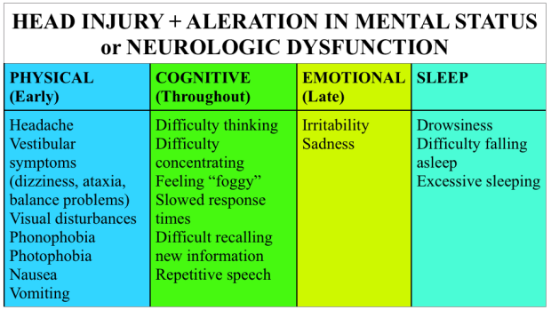
Symptoms: There’s a good bit of overlap with migraine, more significant head trauma, and symptoms that may be confused for behavioral issues in younger populations.

Screen Shot 2015-02-19 at 1.27.09 PM

Testing: Not all head injuries require imaging.  Certainly, patients with focal neurologic findings warrant imaging; yet, in other cases, validated decision aids exist to help determine which patients may or may not need imaging.  In adults, we like the Canadian CT Head tool , although the ACEP clinical policy uses the New Orleans Criteria.  For a solid review of the two, check out this SGEM episode.  In children, consider the use of the PECARN decision aid. Otherwise, a good neurologic exam and observation should suffice.

Something to consider – patients often believe that a “normal” CT scan of the head means that they don’t have a concussion.  As a result, they may not take concussion precautions seriously.  In this case, imaging may provide false reassurance.

There’s increased attention on biomarkers like GFAP, total tau, and S-100B but these are not ready for prime time and are not incredibly specific [3].  Further, standardized assessment tools such as SCAT3 and ACE may be useful, but are used predominantly on-site for sports related incidents.

Treatment

  • Education – this is one of the biggest areas in which ED providers may make a difference. Give patients or family members precautions for concussion, even if the injury or mechanism seems relatively mild.  Studies show that a majority of pediatric patients do not follow up as instructed after a concussion [4].  Perhaps communication of the the potential gravity of concussion and long-term implications may improve follow up.
  • Rest – This the is the mainstay of initial treatment for most mild TBI [1-3, 5].  It’s unclear how strict this rest needs to be, but a recent study in the pediatric literature found no benefit to strict rest versus standard instructions (1-2 days rest + graduated return) [6].
  • Graduated return to activity – after a period of rest, it’s recommended to slowly resume activities, spending at least 24 hours at each level of increased activity.  If an individual gets symptomatic, they should return to the level of activity at which they were asymptomatic (see the Zurich protocol).
  • Return to play – clearing people from the ED is NOT a good idea.  For those on the sidelines, a player should be removed from activity for the day after a suspected concussion.

Why do we care in the ED?

  • Quality of Life – Concussive symptoms can be quite disabling.  Giving patients a name for their symptoms, resources, and education may help them understand the process of recovery and available resources.
  • Second Impact Syndrome – A second head injury in a patient symptomatic from a concussion may experience diffuse cerebral edema, possibly from loss of cerebral autoregulation.* [2]
  • Post Concussion Syndrome (PCS) – a subset of the concussion population will have persistence of symptoms which, if they persist > 1 week to 3 months after the injury, is deemed PCS. *  This is thought to be more prevalent in individuals with a history of depression, anxiety, or migraines and in those with more severe ED presentations [2].
  • Bouncebacks – Patients may present to the ED with symptoms of concussion but may not give a history of significant trauma.  Remember that concussion may occur after a seemingly minor bump to the head and elderly may need extra support with activities of daily living or may bounce back with a more life threatening injury.
  • Lack of follow up.  While some may follow up with concussion specialists, the majority of our patient population lacks the ability for meaningful or specialist follow up.  We can’t assume that someone else is going to guide our patients through concussion recovery or that when we write “follow  up with _____” that it will happen.  Our care may be it, make it good.

*The literature on these is sparse and complicated by variable definitions, information, and standardized reporting.

 Generously Donated Rosh Review Questions (scroll for answers)

Question 1. A 33-year-old man with no past medical history presents with a headache 3 days after a closed head injury. The patient states that he stood up from kneeling and hit the top of his head on a wood cabinet. There was no loss of consciousness or seizure activity. In addition to the headache, he complains of difficulty concentrating at work and dizziness. His physical examination is unremarkable.

Question 2. A 42-year-old man is brought to the ED after he tripped and fell while he and his wife were on a walk. His wife notes that she saw him hit his head on the pavement and that he did not respond to her for 45 seconds. When he started to respond, she says that he was very confused. In the ED, his vital signs are BP 135/75, HR 88, RR 14, and oxygen saturation 98% on room air. On exam, you note some minor lacerations on the patient’s upper extremities, face, and scalp and his GCS is 15. As the wife recounts what happened, the patient does not recall any of the events and continuously asks to repeat what happened to him. A CT scan of the brain is normal. The patient is diagnosed with a concussion and is ready to be discharged from the ED. 

References

1. Tavender EJ, Bosch M, Green S, et al. Quality and consistency of guidelines for the management of mild traumatic brain injury in the emergency department. Acad Emerg Med. 2011;18:(8)880-9. [pubmed]

2. Haydel M.  Management Of Mild Traumatic Brain Injury In The Emergency Department. Emergency Medicine Practice.  September 2012 Volume 14, Number 9

3. Jagoda AS, Bazarian JJ, Bruns JJ, et al. Clinical policy: neuroimaging and decisionmaking in adult mild traumatic brain injury in the acute setting. Ann Emerg Med. 2008;52:(6)714-48. [pubmed]

4.Hwang V, Trickey AW, Lormel C, et al. Are pediatric concussion patients compliant with discharge instructions? J Trauma Acute Care Surg. 2014;77(1):117–22; discussion 122.

5. Brown NJ, Mannix RC, O’Brien MJ, Gostine D, Collins MW, Meehan WP. Effect of cognitive activity level on duration of post-concussion symptoms. Pediatrics. 2014;133(2):e299–304.

6.Thomas DG, Apps JN, Hoffmann RG, McCrea M, Hammeke T. Benefits of strict rest after acute concussion: a randomized controlled trial. Pediatrics. 2015;135:(2)213-23. [pubmed]

Answers

 1.  D.  The patient presents with minor head trauma and complaints consistent with a concussion and should have neurology follow up arranged. A concussion is a minor TBI that is often seen in MVCs and collision sports (football, hockey). It is typically caused by a rotational injury or an acceleration-deceleration injury. Patients will present with a number of non-specific symptoms including headaches, dizziness, confusion, amnesia, difficulty concentrating, and blurry vision but do not have focal neurologic findings. Despite the absence of severe intracranial injury, patients can have chronic and debilitating symptoms from concussions. Neurology referral is recommended, as patients should have functional testing and tracking of their symptoms for resolution. It is vital to counsel patients to avoid contact sports or activities that increased the risk of recurrent injury as these patients are at risk for more severe injury with second impact.In the absence of focal neurologic findings, absence of antiplatelet or anticoagulant use and minor trauma, imaging is not needed (A, B, C).

2. Cerebral concussions are clinically characterized by headaches, confusion, dizziness, and amnesia for the event. Concussions are characterized by a transient loss of consciousness that occurs immediately following blunt, nonprenetrating head trauma, caused by impairment of the reticular activating system. Concussions present without focal neurologic deficits, and CT and MRI show no acute abnormalities. Although not commonly performed for concussions, functional imaging, such a PET scan, may show changes in blood flow and glucose uptake. It is critical to inform the patient and his or her family of the second impact syndrome (SIS), which occurs when a patient suffers a second concussion after being symptom-free from the first. Although SIS is more common in sports based injuries, especially among teenagers, the risk of serious sequelae following a second concussion is immense. Due to neurochemical and autoregulatory changes that may still be present, a second concussion soon after a first generally produces a rapid neurologic decline that proves fatal. Patients should be told to avoid activities that could cause falls or trauma for at least 1 week after the patient is completely asymptomatic from the first concussion. Most patients with a concussion can be discharged from the emergency department and advised to follow-up with a primary care physician within 1 week (B). Although some patients have only transient symptoms from a concussion, others may experience persistent symptoms termed postconcussive syndrome (PCS).  PCS symptoms most often include headache as well as memory, sensory, sleep, and concentration disturbances. It is important to consider PCS in all patients with a concussion, but a primary care physician is generally able to care for these patients. It is unnecessary to obtain an MRI (C).  Although patients should avoid trauma and falls for at least 1 week after being completely asymptomatic after the first concussion, a change insleeping position (D) is unnecessary.

FOAMcastini – Reflections on ACEP tPA Clinical Policy Update Draft

(iTunes or Listen Here)

As detailed in this FOAMcastini, ACEP just released a draft of an updated clinical policy on tPA for acute ischemic stroke.  This came in the wake of years of controversy over the aggressive position taken in the 2012 clinical policy.

While FOAMcast is not an interview style podcast, we felt compelled to get some perspective on Emergency Physicians a little more experienced than ourselves.  Here we interview:

Dr. Ryan Radecki (@emlitofnote), Assistant Professor, University of Texas – Houston

  • We don’t know who best benefits from tPA so elucidating which patients are “carefully selected” may get hard.
  • See his response to the policy on his blog here

Dr. David Newman (#draftnewman), Associate Professor of Emergency Medicine, Mount Sinai Hospital

  • The process for ACEP clinical policy creation seems to work.  The constituency expressed concern and the college listened and went back and re-created the policy from the bottom up.
  • This policy reflects a move from content expert to methodologists which better reflects the evidence compared with opinions (and is the standard per USPSTF).

Dr. Anand Swaminathan (@EMSwami), Assistant Professor of Emergency Medicine, NYU

  • Inclusion of more rigorous methodology and review of evidence.
  • May be perceived as too “soft” by tPA supporters.  This policy may not overtly change practice but may open up avenue of conversations.

Dr. Ken Milne (@thesgem), Chief of Staff at South Huron Hospital

  • When looking at “carefully selected” patients, as noted in the policy, remember to use the Evidence Based Medicine trifecta of evidence, patient values, and clinical expertise.  In isolation, one component is not sufficient.
  • Previous discussions of NINDS, and the Cochrane tPA article

Episode 16 – Headaches

(ITUNES OR LISTEN HERE)

The Free Open Access Medical Education (FOAM)

This week we review a post from Dr. Rob Orman’s ERCast, Is it really a sinus headache?

POUND- 4 criteria is very indicative of migraine (+LR 24), 3 criteria also likely (+LR 3), although most of this comes from the outpatient literature [1].

  • Pounding headache
  • hOurs: headache lasts 4-72 h without medication
  • Unilateral headaches
  • Nausea
  • Disabling: disrupts daily activities

The Bread and Butter

We summarize some key topics from the following readings, Tintinalli (7e) Chapter 159 ; Rosen’s 8(e) Chapter 20, 103 – but, the point isn’t to just take our word for it.  Go enrich your fundamental understanding yourself!

In Emergency Medicine, our job is to investigate and think about the life and limb threatening causes, even to mundane problems.   Things such as intracranial bleeds, meningitis, masses – these are huge deals and are covered well and hammered into our heads.  For FOAM core content on this, check out the St. Emlyn’s podcast.  On this episode, we’re running a mini-ophthalmology headache special and focusing on headaches that treatment may render “sight saving.”

Temporal Arteritis – often in patients older than 50 years of age and more common in those with a history of polymyalgia rheumatica. May be accompanied by visual changes including the “classic” amaurosis fugax or “curtain” of unilateral vision loss.  If not treated, these patient can lose vision permanently.

  • Unilateral or localized headache, often in the temporal or retro-orbital area
  • Jaw claudication (pain with chewing) – most specific sign
  • Decreased pulse in temporal artery or tenderness
  • Sedimentation Rate (ESR) >50

Treatment

  • Prednisone 40-60 mg if thinking about diagnosis
  • Temporal artery biopsy within 48 hrs

Acute Angle Closure Glaucoma – Classically, these patients present with unilateral mid-dilated pupils and severe nausea, vomiting, and headaches.  The history can, naturally, be less classic and more vague.  Also, if not treated, this can lead to vision loss.

  • Elevated intraocular pressure (>20 mmHg)
  • Decreased visual acuity
  • Fixed irregular semidilated (midposition) pupil
  • Slit lamp — shallow AC (closed angle), injected conjunctiva; corneal microcystic edema (cloudy)

Treatment –

  • Ophthalmology consult stat
    • They may want topical b-blocker, cholinergic, alpha-2 agonist, eye drops or administration of acetazolamide

Idiopathic Intracranial Hypertension (Pseudotumor Cerebri) – Common in young, overweight women or those on oral contraceptives.  Untreated, they can suffer vision loss.

  • Elevated opening pressure (>20-25 cm H20) on lumbar puncture

Treatment

  • Neuro follow up
  • Acetazolamide +/- furosemide
  • Therapeutic lumbar punctures

Cerebral Venous Sinus Thrombosis – may present as atypical headache with stroke like symptoms in patients without known vascular risk factors.  The neurological findings may be transient.  Often associated with post-partum patients, patients with hypercoaguable states (Factor V mutations, protein C or S deficiency, antithrombin III deficiency, etc), patients on OCPs.

Diagnosis – CTV or MRV (magnetic resonance venography) after CT scan, which may be normal.

Treatment – Anticoagulation, although this is somewhat controversial

Generously Donated Rosh Review Questions (Scroll for Answers)
Question 1. A 73-year-old woman with a history of hypertension presents with a unilateral headache for 3 weeks. She states that she has a throbbing pain at her right temple and has pain in her jaw with opening and closing. The vision in her right eye has worsened over the previous day. Her blood pressure is 173/100.
Question 2. A 71-year-old woman presents to the ED with daily headaches for 2 months. She describes the headache as a dull pain that is most intense in the morning and resolves by the afternoon. On exam you note 4/5 motor weakness of the left upper and lower extremity.
References:
1. Detsky ME,McDonald DR, Baerlocher MO, Tomlinson GA,McCrory DC, Booth CM. Does this patient with headache have a migraine or need neuroimaging? JAMA. 2006 Sep 13;296(10):1274-83.

2. Chapter 20, 103.  Rosen’s Emergency Medicine, 8e.

3.Chapter 159.  Tintinalli’s Emergency Medicine: A Comprehensive Study Guide, 7e. New York, NY: McGraw-Hill; 2011

Answers
1. D. This patient presents with a unilateral, subacute headache with associated jaw claudication and vision change; symptoms consistent with temporal arteritis. Temporal arteritis or giant cell arteritis is a systemic inflammatory process of small and medium-size arteries. The most commonly involved vessels are the ophthalmic vessels and the extracranial branches of the aortic arch. The disease typically affects patients over 70 years of age and is more common in women than in men. Patients present with a subacute headache that is throbbing in nature and may be present for weeks to months. Often, patients will have symptoms for more than 2 months. Patients may also report jaw claudication secondary to vascular insufficiency of the masseter and temporalis muscles. Physical examination may reveal tenderness over the temporal artery. Systemic symptoms may also be present including fever, joint pains, and weight loss. Diagnostic testing in the Emergency Department generally begins with an erythrocyte sedimentation rate (ESR) with a cutoff of 50 mm/hour although the level may be >100 mm/hour. However, the ESR will be normal in 10-25% of patients. The gold standard diagnostic test is a temporal artery biopsy. In patients with a high-clinical likelihood of temporal arteritis, treatment should be initiated regardless of initial diagnostic testing as delay can lead to permanent visual loss. Prednisone should be started at 60 – 120 mg/day.

Carbamazepine (A) is the treatment of choice for trigeminal neuralgia, not temporal arteritis. The patient does not present with symptoms consistent with hypertensive emergency requiring emergent antihypertensive treatment withlabetalol (B). A non-contrast head CT scan (C) is not helpful in temporal arteritis as the disease does not involve the intracranial contents.

2.  B  More than half of patients diagnosed with a brain tumor complain of headache. However, the headache associated with brain tumor is highly variable. Patients may describe it as continuous or intermittent, unilateral or bilateral, sharp or dull. It is associated with neurologic deficits less than 10% of the time. However, in the setting of aneurologic deficit and chronic headache (as in this scenario with motor weakness), a mass lesion should be strongly considered as the cause. Patients may also complain of nausea, vomiting, visual change, and gait disturbance. Headaches due to brain tumors are classically associated with pain that is worse in the morning (as in this case). However, this is rare.

Central venous thrombosis (A) results from hypercoagulable states and is associated with acute to subacute headaches with vomiting and sometimes seizures. Risk factors include the use of oral contraceptives, postpartum or postoperative states, and any hypercoagulable state such as factor V Leiden mutation, antithrombin III deficiency, protein S or C deficiency, or polycythemia. The diagnosis is usually made by MRI venogram. Migraine headache (C) is classified as a primary headache and can be quite variable in presentation. These headaches can be associated with nausea, vomiting, photophobia, and phonophobia. The headache may also be preceded or accompanied by an aura that develops gradually over minutes, usually lasts 60 minutes, and is reversible. Auras may include neurologic symptom but commonly include scintillating scotomas (dark spots) or flashing lights. Temporal arteritis (D) occurs almost exclusively in patients older than 50 years and is much more common in women. Headache is the most common symptom of temporal arteritis and usually occurs over the frontotemporal region. It is strongly associated with a history of polymyalgia rheumatic. It is not associated with focal neurologic deficits, but it can lead to vision loss due to ischemic optic neuritis.